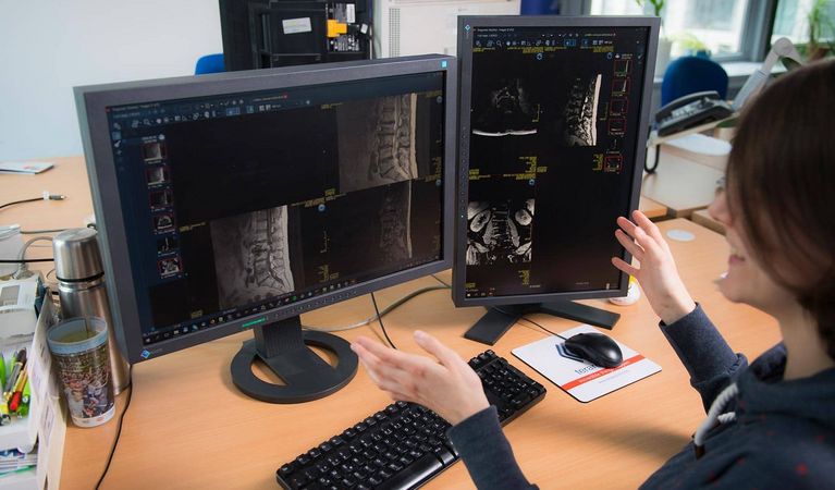
The term soft tissue tumors covers tumors of various tissues (musculature, connective tissue, fatty tissue, nervous tissue). An important part of their diagnosis as well as the determination of a successful therapy is the correct assessment of the tumors on medical scans. To do this, medical professionals visually analyze the tumor on MR, CT and PET scans. These images thereby capture the tumor in different anatomical, functional and molecular contexts, providing different information about the tumor. Depending on the clinical question - for example, is it a matter of biopsy, radiotherapy or surgical planning? - different aspects and images are relevant.
Deep Learning makes segmentation more precise
Until now, the segmentation of tumors has been a manual and time-consuming task that takes up a lot of radiologists' attention. Automated tools can be a valuable addition in everyday clinical practice. In the strategic research project Integrative Visual Computing and as part of the FEMtech internship Tumor Segmentation precisely these challenges were addressed. FEMtech intern Theresa Neubauer, under the supervision of Biomedical Image Informatics leader Katja Bühler, researcher Maria Wimmer and with cooperation partner Thomas Beyer from MedUni Vienna researched solutions to improve the segmentation of soft tissue tumors on multimodal images. Until now, the potential of multimodal data has been exploited by a few established segmentation methods. Therefore, VRVis developed a new method that combines multimodal information from different imaging modalities. For this purpose, MR, CT and positron emission tomography data are fused to obtain a more accurate view of the tumor and to segment it more precisely. The new method relies on machine learning, which learns all the multimodal features for the tumor segmentation task at once. This not only speeds up the process, but also makes it more accurate. The research work and specifically the internship also resulted in a scientific paper published at the prestigious MICCAI conference (International Conference on Medical Image Computing and Computer Assisted Intervention).
AI supports the digital radiology of the future
Accelerating everyday medical practice with the help of artificial intelligence is a major goal of digital radiology. VRVis researches solutions towards this goal for over 20 years. The aim is to facilitate the diagnostic work of physicians while always keeping the focus on people and keeping the human in the loop ("human-centered AI"). The machine provides a valuable basis for decision-making, while doctors continue to make the decisions themselves. This speeds up radiological workflows in hospitals and helps to save more lives.
More information
September 21, 2021