

Images hold a lot of information and are a universal human language. For over 20 years, VRVis has been researching how computers can identify, classify, interpret, and analyze the content and information from these images as efficiently as humans, if not even more so.
Our primary methods in the field of image analysis, computer vision, and video processing are based on conventional image analysis topics, i.e., machine and deep learning for classification, segmentation, object detection, and annotation of image and video data, on image correlation, as well as on visualization of image data.
We develop tailor-made methods in collaboration with our research partners and customers according to the characteristics of the image data and tasks in each case to implement specific solutions for image-based decision-making.
To support complex, transparent decision-making processes, we conduct research into innovative ways of enabling not only image information recognition but also visualization and further networking of decision-making procedure. Our automated, image-based search, analysis, and recognition methods support, accelerate or replace human decision-making processes in medicine, industry, and science.
Today, rapid technological developments in imaging methods enable better insights, for example, into biological processes. Methods for analysing and correlating image data from different sensor sources and image modalities, and the visualization of this data, which is often immense and heterogeneous, are a key technique in interpretation and are being researched and developed by us in collaboration with imaging specialists. VRVis is a member of the Austrian BioImaging/CMI network and thus has access to high-end correlative imaging technology and expertise.
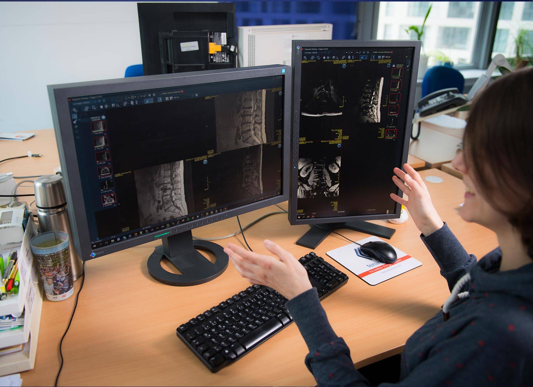
Digital radiology
VRVis has been researching solutions for digital radiology for more than 20 years. Our innovations support medical teams in disease prevention, pathology detection and therapy planning. The results of our research have been published several times and patented internationally. Our portfolio of solutions and digital tools ranges from fully automated semantic annotations, cardiac analysis, AI-assisted tuberculosis screening to soft tissue tumor segmentation in multimodal data or trustworthy AI solutions (XAI). More information about our digital radiology solutions.

Medicine
Using the latest methods of visualization, image processing and data science, we accelerate medical diagnostic processes, surgical planning and intervention planning for our partners in the biotech industry and healthcare sector. With our data platforms and data science solutions, we contribute to a better understanding of epidemiological processes and the development of precision medicine. More information about our applications and technologies for medicine.

Neuroscience
State-of-the-art imaging techniques and progress in the development of genetic tools allow ever deeper insights into the functioning of nerve cells, neural networks and the entire brain. In collaboration with renowned international institutions, VRVis has been developing data science methods for the computational analysis of neuroscientific data as well as big data platforms for the management, integration, visualization and, above all, mining of large collections of neuroscientific image and network data and their molecular context for over a decade. More information about our solutions for computational neuroscience.

Visualization
The correct handling of multidimensional, multi-channel and/or time-dependent image data as well as entire image collections is increasingly becoming a bottleneck in data analysis. Visualization is an important tool to fully exploit the wealth of information from image data. This includes the visualization and interactive analysis of brain network data, high-performance 2D and 3D visualization of brain data and visualization methods for behavioural biology. More information about visualization solutions for neuroscience and life science.

Materials science and manufacturing
Modern materials science and production companies use image processing methods to evaluate data from sensors and cameras. This supports production processes and optimizes quality inspections. At VRVis, we develop AI-based image processing methods for our partners such as RHI Magnesita to automate visual quality inspections and analyze material structures for damage types as well as to simplify annotation for future training data.

Satellite image processing
Satellite data from various missions and programs provide a wide range of data, but must first be processed in order to access the information they contain. Digital image processing is therefore one of the key technologies in remote sensing. VRVis develops image processing methods to obtain relevant information for agriculture from satellite images. This includes, for example, the size and structure of agricultural areas. We also use super-resolution reconstruction (SRR) to improve the resolution of image data with AI and thus generate even more detailed information, such as vegetation characteristics. More information about this project.

Pests and extreme weather affect grain growth and yields. Recording and analysing plant development provides the basis for the prevention of crop losses. VRVis is part of a project consortium developing a platform for agriculture with the aim of using functional imaging data for analysis and teaching skills in training courses.
VRVis is supporting Hage Sondermaschinenbau with its expertise in machine learning: Hage needs a monitoring system for spacious facilities to ensure that no people are within the machines' activity range. Thus, VRVis is testing the suitability of ML methods for safety in industrial environments.
Regular maintenance of the vehicles is necessary for safety in rail transport. When working on the train roof, technical personal might forget spare parts or tools. This can be dangerous when going at high speed. VRVis and Zugkraft-kN are designing a scanner for train roofs, automatically detecting this problem.
With the help of 3D printing (additive manufacturing), spare parts for defective trains can be produced more easily and in a sustainable way as well as faster and cheaper - a great potential for the climate-friendly future of train transport companies.
The aim of the application project IVC Multi is to research novel intelligent visual computing methods supporting decision-making in automotive industry, medicine, and life sciences based on ensembles of heterogeneous, multi-scale and/or multi-temporal data.
The aim of the project IVC Stream is to research novel visual computing solutions for simulation and measurement data.
This project aims at accelerating and automating image-based decision making with an application focus on medicine, recycling and quality assurance processes in manufacturing.
The strategic project of the area is the organizational and scientific hub for the realization of the area-wide intelligent visual computing approach for analytics and modelling based on ensembles of dense grid-based data, derived data, and digital embedding.
Within the project Larvalbrain 2.0, a dynamic multi-scale multi-level atlas and data collection of structural, molecular, physiological, and behavioral results of Drosophila melanogaster larvae will be established.
VRVis a founding member of the Austrian BioImaging/CMI, which is a professional consortium of multiple Austrian science institutions and the official Austrian Euro-BioImaging initiative.
COMULIS is an EU-funded COST Action that aims at fueling collaborations in the field of correlated multimodal imaging (CMI).
The INDIGO research team is systematically documenting the graffiti on Vienna's Donaukanal, using these photos to create a digital twin of the walls. VRVis is currently contributing its technological expertise to the project.
Visual computing for medicine: image processing solutions for new applications in radiology.
In this project we develop new methods from the field of visual analysis and machine learning to automate the quality control and quality assurance of glass articles.
The long-term vision of this applied research project is to use available data resources to improve image-based diagnostics based on complex data in daily clinical routine.
The strategic project forms the organizational and scientific hub for the realization of an area wide integrative visual computing approach. It covers joint strategic research and development on fundamental challenges in all application projects.
For centuries, neuroscientists have been mapping the brain. Until now, the step from simple maps to a generally accepted model has proved to be extremely difficult. In this project, a 4D atlas of the brain of the fruit fly larva is being built.
Visual computing techniques for the automated detection of osteoporosis and osteoarthritis.
Software for the use of multi-modality images in external radiotherapy.
Next generation workflows for interactive knowledge generation from images and simulations.
The analysis, visualization and exploration of high-dimensional image spaces are the subject of the KAFus project.
To enable people to live an economically, ecologically and socially sustainable life, applications by VRVis contribute to 10 of the 17 Sustainable Development Goals (SDGs). Due to this, VRVis is now nominated for the Austrian SDG-Award.
VRVis was invited to present its workflow for a digital, interactive exhibition archive for Schloss Trautenfels in this year´s IÖB Challenge.
New VRVis AI method that boosts confidence in computer-aided diagnosis receives the eAward 2021.
On March 3, 2020, Katja Bühler, head of our Biomedical Image Informatics Group, was awarded with the renowned TU Women's Prize.
Florian Ganglberger's short paper received an Honorable Mention at the EG VCBM 2019.
Visual computing for computer-aided diagnostics and operation planning.
A. Neubauer, M. T. Forster, L. Mroz, R. Wegenkittl, K. Bühler, STEPS - An Application for Simulation of Transsphenoidal Endonasal Pituitary Surgery, in Proceedings of IEEE Visualization 2004, pp 513-520. 2004, IEEE Vis 2004 Best Applications Paper
The European project SUMMER won the award 'Les étoiles de l'Europe'.
Sebastian Zambal, Jiří Hladůvka, Armin Kanitsar, Katja Bühler, Shape and Appearance Models for Automatic Coronary Artery Tracking, WON MICCAI 2008 Contest: 3D Segmentation in Clinic: A Grand Challange.
J. Beyer, M. Hadwiger, S. Wolfsberger), K. Bühler, High-Quality Multimodal Volume Rendering for Preoperative Planning of Neurosurgical Interventions, in IEEE Transactions on Visualization and Computer Graphics 13(6) pp.1696-1703 / Proceedings of IEEE Vis 2007, Vis 2007 Best Application Paper
A. Neubauer, Endoscopy for Preoperative Planning and Training of Endonasal Transsphenoidal Pituitary Surgery.
C. Langer, M. Hadwiger, K. Bühler, Interaktive diffusionsbasierte Segmentierung von Volumendaten auf Grafikhardware, Bildverarbeitung für die Medizin 2005; GI Informatik Aktuell; Springer Verlag. pp 168-17, BVM 2005 Best Poster
S. Wolfsberger, M. Donat, A. Neubauer, K. Bühler, T. Czech, E. Knosp, Virtuelle Endoskopie in der transsphenoidalen Hypophysenchirurgie, CURAC 2005 Best Poster
M. Meissner, B. Lorensen, K. Zuiderveld, V. Simha, R. Wegenkittl, Volume Rendering in Medical Applications: We've Got Pretty Images, What's Left to Do?, in IEEE Visualization 2002